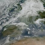
Spectral Cognisance 3 – When Black turns Red

We have established that emission and absorption spectra are quite important in image sensing.

Why is this so?

First, an image sensor cannot distinguish between wavelengths. All that matters is the number of photons. There are, however, some more subtle effects.

  • Color sensors can distinguish wavelengths.
  • Different media have different sensitivity to wavelengths, i.e. image sensor A vs. B, human eyes, film… I will discuss some of this in the 4th post in this series.
  • Blue photons deliver more energy and have a shallower absorption depth than “red” photons. This will make a difference in a later post on Anti-Blooming and Shutter Efficiency.

Today, I will focus on the color reproduction of a silicon sensor under certain spectral emission content. I have been in imaging long enough to see the advent of mainstream machine vision color image sensors, readily available from all major camera vendors – including us. Coming from a largely black-and-white world, there were a few things that needed to change when applying color.

Return once again to our spectral plot. This time I changed the monochromatic sensor absorption curve (QE) to a typical response curve with RGB (Red-Green-Blue) filters on the array.

Figure 1:  RGB Sensor Response and Black Body Emission (3500K)

There are two point sI want to consider here:

1) You can clearly see that there will be many more photons passing the Red filter than the Blue filter, basically shown by the overlapping areas of Emission and QE curves.

2) If you look closely, you can also see that many photons will pass ALL filters to different degrees for different wavelengths. This is most pronounced for wavelengths above ~700nm.

The first point – different “available” intensities for different colours – results in a certain color balance of the image. Reds and yellows will be more pronounced than blues. As humans, having lived with sun light and light bulbs for a long time, an image balanced thus will still appear “white”. There is one caveat: an image sensor tends to be less responsive in blue than in red, so the incoming blue signal is further reduced. Therefore, “white balancing” (meaning making a digital image look white) usually increases the “blue gain” by approx. 2x over the red to accommodate not for the lighting but the non-uniform sensor response.

The second point was of major interest for me when we developed our first colour camera.

A Field test with a new camera delivered the following image.

Red Hat shwoing with Standard Coloir Imager

Figure 2: Standard Color Image

At first glance, it seemed nothing was wrong with this image. Except… weren’t those old DALSA caps black???

Black Hat showing with IR-cut Imager

Figure 3: Color Image with IR-cut Filter

Yes indeed, they were!

What happened?

The image in Fig 2 was taken with the camera “as-is”, meaning the sun’s black body emission (~5000K) is allowed to create photons in the RGB sensor at will. Review what we discussed in Fig 1 and you will see that there are a lot of red photons that appear in the image as indistinguishable color (they pass through all R, G and B filters similarly). Hence black, where there are no photons in the visible spectrum, can still reflect deep red photons (>700nm) which in turn create signal on all filters with the R filter showing the dominant signal. The black cap turns red.

In addition, if you look at the plants in the foreground, you can see that some of the green/yellow information is distorted. The plant seems to reflect some of the deep-red spectrum where all RGB filters transmit and distort the nice green to a brownish yellow.

Once we add an IR-cut filter in Fig. 3, effectively removing all spectral content above ~650 nm for the sensor we obtain a nice image with good color reproduction. A next step is to add color processing to the data stream, to achieve the highest possible color fidelity.

This raw image shows, however, that the information content from the sensor is quite decent when an appropriate spectral content is offered to the sensor. And this is right along the lines of discussion for this series.

Next I will detail a bit more on the color perception of humans in “Is Lux short for Luxury?”

Until then,

Matthias

Matthias

About Matthias

Born in the early seventies in Northern Germany I graduated in Physics at the University of Ulm. I worked for the BOSCH automotive group in Stuttgart before joining DALSA in 2000. Since then I have worked on layout, design, project lead, architecture and program management for various DALSA CMOS image sensors. I enjoy creating sensors and seeing them through production and implementation at the customer end.
Posted on by Matthias. This entry was posted in CMOS, Image Sensors. Bookmark the permalink.

4 Responses to "Spectral Cognisance 3 – When Black turns Red"

  1. Pingback: Image Sensors: When Black turns Red, by Matthias at Teledyne DALSA | Digital Cinema Tools | Scoop.it

  2. Pingback: Image Sensors: When Black turns Red, by Matthias at Teledyne DALSA | Visual Culture and Communication | Scoop.it

  3. In the last years I have encountered this phenomen several times. I’m speaking mainly of the red cast in the black cap that I believe may be or not connected with the general color cast issue (reddish plants). I have noticed that the phenomenon was particularly strong on specific materials, like synthetic tissues (i.e. typically on CD folders, socks, etc.). Finally I have come to the conlusion that this is caused by some sort of band shift (incident visible light reflects some IR on some specific materials). I have come to this conclusions because I have noticed this phenomen using several light sources (i.e. White LED) with no IR emission in combination with different IR CUT OFF Filters. Most CCD I use are sensitive up to 1050nm, while most of the IR CUT-OFF filters were very effecttive removing IR until 950-1000nm only but than they slightly “re-opened” after that, leaving a small interval in which the sensor could possibly see IR. I tryed different IR CUT OFF filters (from 650nm to 750nm) without being able to completely remove the red cast in these special materials, until I found some filter that did not re-open at all in these intervals, and I was finally free from the red cast in the black synthetic materials ! But from where did the 1000nm IR light came from (I’m sure my light source was not emitting it) ? The only explaination to me was a band-shift effect (sort of IR inverse fluorescence effect) caused by these materials. Does this make sense to you ? Do you have a different interpretation ?
    Another issue I discovered (concerning general color accuracy) is that IR CUT filter tend to close to IR too quickly; but because this is done at the edge of human vision it cause a color accuracy problem and reddish cast. Le’ts select a 750nm IR filter for an example: Human vision drop softly in the RED range and sensitivity is very low close to the limits, while a CUT OFF filter will allow a lot of RED photons to pass until the CUT-OFF limit. This cause a steep edge and a reddish cast on most images. Selecting a different CUT-OFF filter (i.e. 650nm) will remove more RED but will also cause the loss of some information on some specific colors, degrading color accuracy anyway. A standard BG-38 like filter will generally provide a much better color accuracy respect to IR-CUT off because of the smoother transition in the RED-IR interval which is much closer to human eyes (but than there other issues as those filters do not block IR very effectively and have many other limitations). So in fact up to now I could not find the perfect filter capable of providing the best possible color accuracy, because either I’m dealing with red color casts (IR CUT OFF above 700nm), either I’m loosing some red colors (IR CUTOFF<700nm), either I have IR coming into the equation (BG-38 like filters). What is your experience on this ? Can you add some information or suggestion ?
    Best Regards

  4. Matthias Matthias says:

    Dear Massimo,
    thank you for your interest and feedback!
    Let me try to respond to your points as orderly as I can manage.

    1) You are seeing IR transmission (and absorption by the sensor) around 1000nm with certain filters, but are not sure where the light is coming from originally.
    1.a) You are completely correct to inspect filters across the full spectral band of silicon (in this case). Indeed many optical filters “open up” significantly outside their specified range, which can indeed cause the same issues – if at lower intensity – as not having a filter at all.
    1.b) To get to the bottom of your question of origin of the IR light I can make the following suggestions.
    Are you studying this effect in a truly dark environment, with your LED (or other known spectral source) being truly the only light source? If you have daylight (tungsten, fluorescent, …) creeping in you may quickly accumulate IR intensity.
    Are you working at very low light levels over all? The colour accuracy of your known light source will be higher if it’s absorbed intensity (after filter, QE, etc.) is substantially higher than the background (if there is any).

    Looking around a bit there are certainly enough materials and dies that fluoresce at longer wavelength. Getting enough intensity together at ~1000nm to provide significant impact on a visible spectrum will be hard. Let’s make the following assumptions:
    - we need a 10% signal intensity from the IR to distort the visible
    - relative filter transmission (1000nm vs. 500nm) is 10%
    - relative QE of 10% (~5% at 1000nm, 50% at 500nm)
    You would need 10x more 1000nm photons than 500nm photons to get this distortion (10% IR = X *10%T*10%QE). Obviously, if you get the visible distorted by 1% IR you need “only” an equal numbers of photons.
    This seems like an unlikely issue to me – the “glowing” plastics would have to have some really specific properties to generate this kind of light power…

    2) You are wondering about reproduction of human colour perception with image sensors.
    I have been looking at this issue as well, not so much from the application side but more from a scientific interest. It baffled me how sensor manufacturers prefer to proclaim their sensor performance using Lux as unit. On closer analysis I came to similar conclusions as you. There is simply no good, clean way to generate a true Lux response from a silicon sensor. Filters do come pretty close and may generally be good enough, but it makes comparing Lux based numbers very difficult. Vendors usually do not specify how exactly they did the tests.
    Incidentally, I am writing the fourth post in this series precisely on these Lux grumblings and my conclusions.
    I am expecting it will come out early next year.
    Why don’t you look in once in a while and we can discuss again later?

    I am open for further discussion if you wish to follow up as well.

    Thanks for your interest,
    Matthias


Leave a Reply

Your email address will not be published. Required fields are marked *

You may use these HTML tags and attributes: <a href="" title=""> <abbr title=""> <acronym title=""> <b> <blockquote cite=""> <cite> <code> <del datetime=""> <em> <i> <q cite=""> <strike> <strong>